
Ventriculites fistulosus
Schrammen 1912
Ventriculites fistulosus is a rare species at Misburg, contrary to V. radiatus. It differs from the latter species mainly by the more irregular shape and arrangement of the pores.

Ventriculites radiatus
Mantell 1822
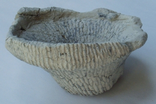
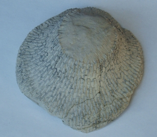
Ventriculites radiatus is a well known species, also from other European localities. Its shape can vary from tubular, as on the left, to flaring funnel-shaped, to virtually sheet-like (below), but its typical pore structure always remains the same. On the outside of the tube wall, the pores (ostia) are oval, two to several mm long, and are arranged "in quincunx", i.e. like the five on a dice. On the inside, the oscules are more or less round and in quincunx.

A nearly complete example of Ventriculites radiatus.

Top view of a funnel-shaped specimen of ? Ventriculites radiatus.
It differs from typical representatives, because the long axes of the postica on the visible inner (upper) surface also show a radial, not tangential orientation. Also, the pores are smaller and the wall thickness is less than in typical Ventriculites radiatus specimens.

Bottom view of a sheet-like fragment of Ventriculites radiatus.

Fragment of Ventriculites radiatus.
top left : underside
top right : upper side (mirror-image)
bottom left : underside, etched
botom right : upper side, etched (mirror-image)
Notice that the long axis of the oval pores is oriented radially on the under side (ostia) and tangentially on the upper surface (postica). Both postica and ostia end blindly. Their bottoms are formed by the skeletal bridges between the pores.

Pore structure of Ventriculites radiatus.
Left: Outer surface of etched specimen, showing regular pattern of large oval ostia separated by microporous cortex.
Right: Detail of microporous cortex. The faint white streaks are rays of cortical lychnisk spicules shining through. The thin siliceous cortex sheets, which make up the inner and outer surface of the sponge, spread out between, and are derived from these lychnisk arms.

View from inside the sponge wall through an exhalent pore of Ventriculites radiatus.
The image shows parenchymal skeleton in the periphery and a dense "furry" lining in the apochete (exhalent canal) made up of the branched ends of parenchymal lychnisks.

Detail of parenchymal skeleton of Ventriculites radiatus, with fused lychnisks with fairly smooth arms and typical octahedral lanterns in their centers.
Notice branching terminations of lychnisks in the lower left corner, similar to those in the pore lining in the picture above.

Detail of skeleton of Ventriculites radiatus, showing parenchymal lychnisks in the foreground and cortex in the background.
Notice transition from the lychnisk arms into siliceous cortical membrane (top left corner).

Detail of skeleton of Ventriculites radiatus, showing very slender, barbed lychnisks and other undefined spicule shapes. Spicules of this type occur locally within the parenchymal skeleton.

Ventriculites stellatus
Schrammen 1902
Fragments of Ventriculites stellatus are moderately common in the Lower Campanian of Höver, but good specimens are absolutely rare.
Typically, Ventriculites stellatus has an obconical shape. The beautiful surface ornamentation, consisting of longitudinally arranged, star-shaped bridges between the ostia, is unique to Ventriculites stellatus. The star ornamentation grades into pronounced longitudinal stripes near the base of the funnel. A stem is not developed. The funnel base rather sits on a set of strong, laterally radiating roots.
The specimen shows a broad (10 mm) growth margin and also two growth rings, probably indicative of stress periods. The walls of the sponge are fairly thick, from 8 to 10 mm.

Another Example of Ventriculites stellatus.

The image shows a wall fragment of Ventriculites stellatus from the gastral (left) and the dermal (right) side. Notice that the postica are oval and arranged in longitudinal rows.


Ventriculites sp
The specimen shown here in two images was earlier thought to belong to Ventriculites stellatus. However, althought it is certainly a Ventriculites sp. species, the star pattern is restricted to the exhalent pores on the gastral side. It is probably closer to Ventriculites successor.
Ventriculites successor
Schrammen 1924
Ventriculites successor is distinguished by the slit-shaped pores on the inside surface, whereas the pores of Ventriculites radiatus are round to oval. Schrammen (1924) considers V. successor as a late mutation, occurring in the upper Campanian, while V. radiatus appears already during the Turonian.
The two pictures show a funnel-shaped fragment in side-view and from below.

Side view of a specimen of Ventriculites successor.

Ventriculites successor with broken and dislocated stem.

This example of Ventriculites successor shows an ontogenetic stage where the sponge was just beginning to spread out sideways and to develop an umbrella-like top.