Cretaceous sponges from the Campanian of Misburg and Höver


Polyopesia angustata

Schrammen 1902

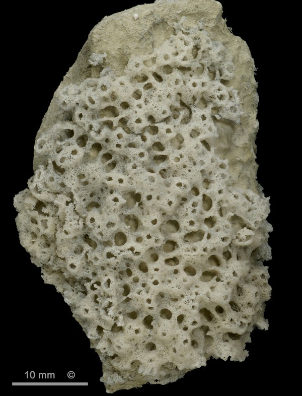
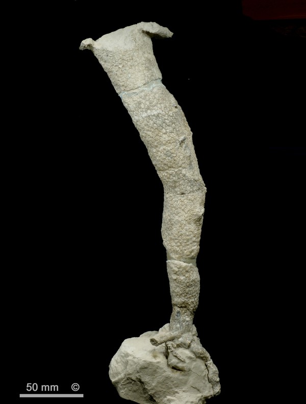
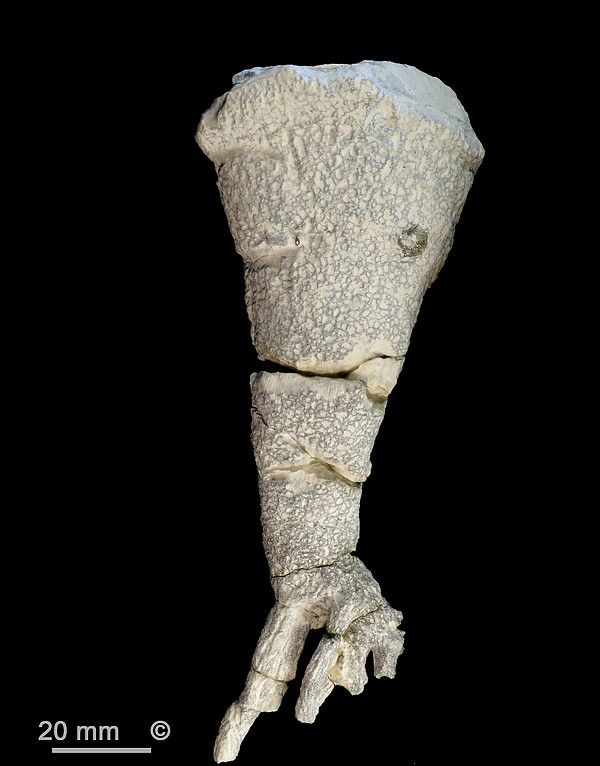
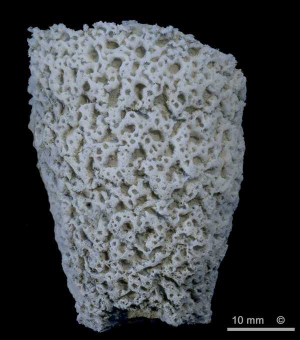
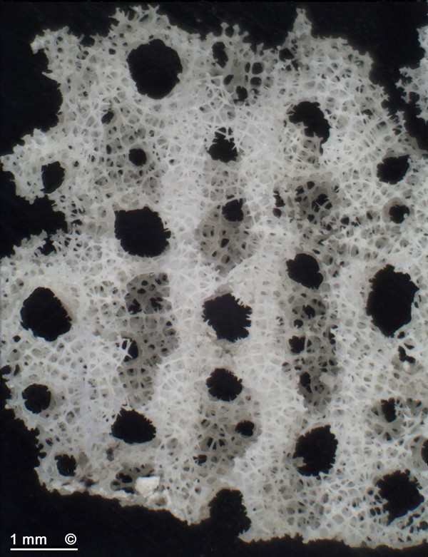
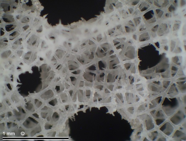
Figure 1 - Polyopesia angustata. Teutonia Nord, Misburg.


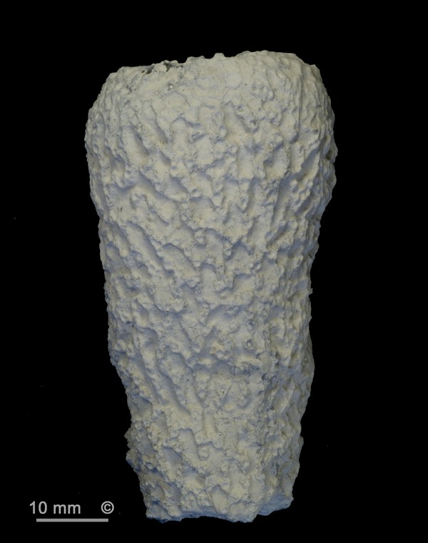
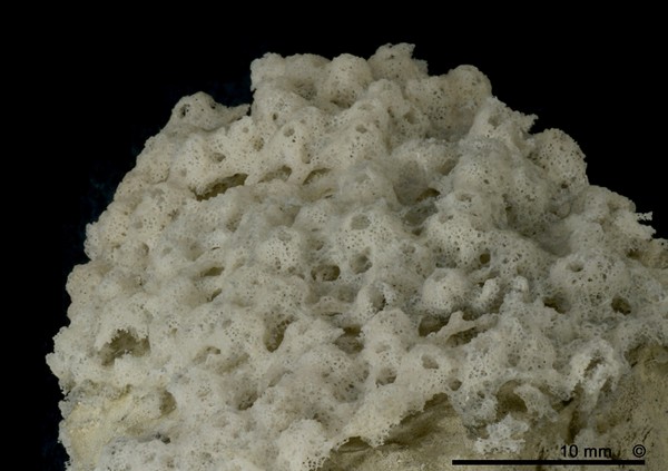
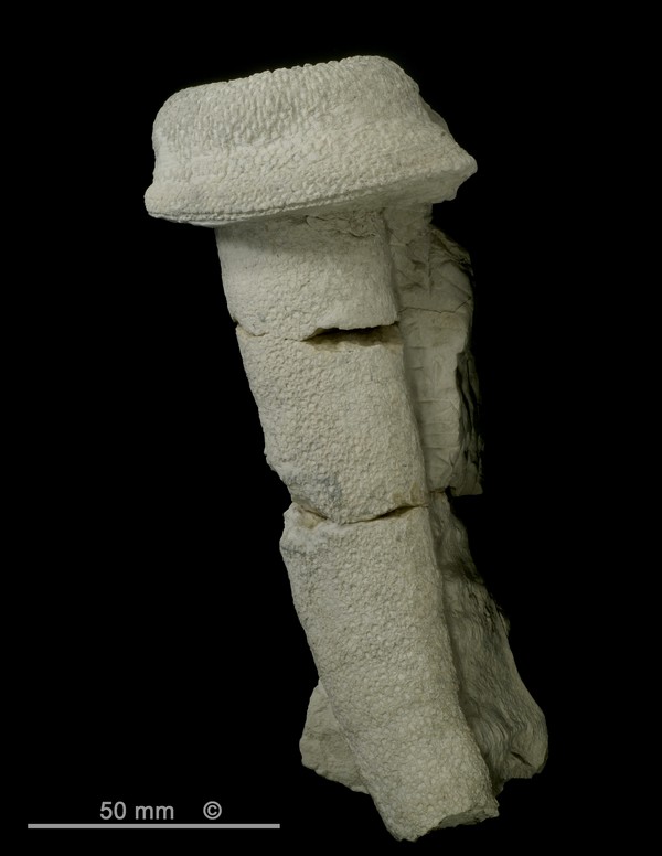
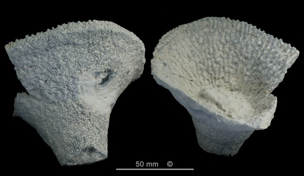
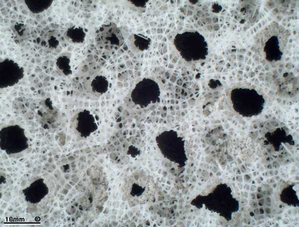
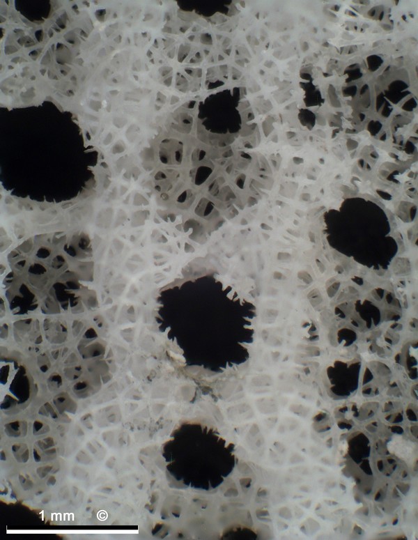
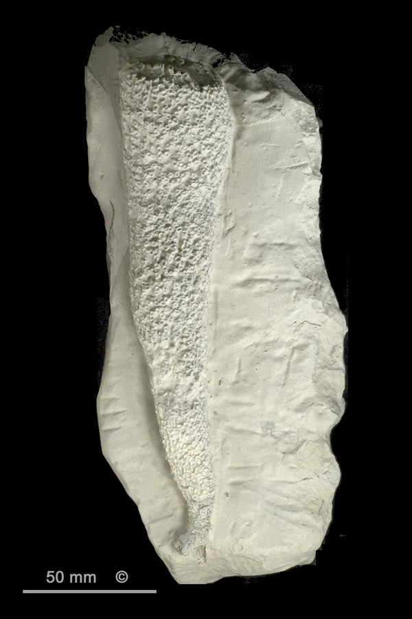
Figure 2 - Polyopesia angustata. Alemannia, Höver.
Top section with constricted growth margin.


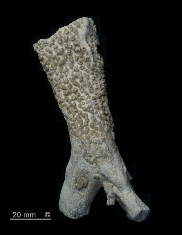
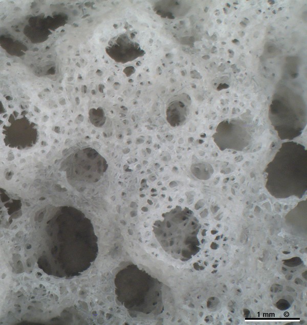
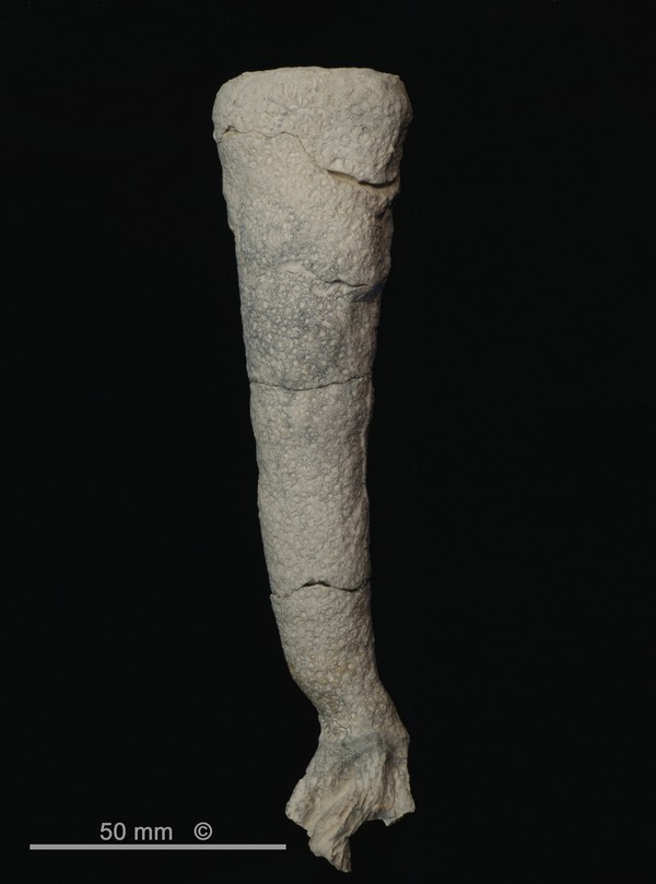
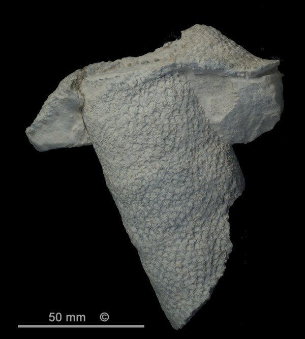
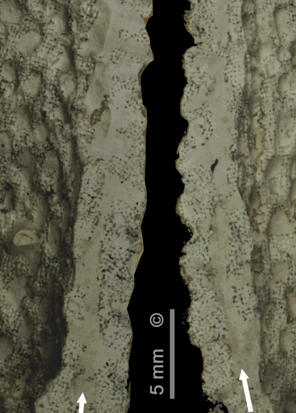
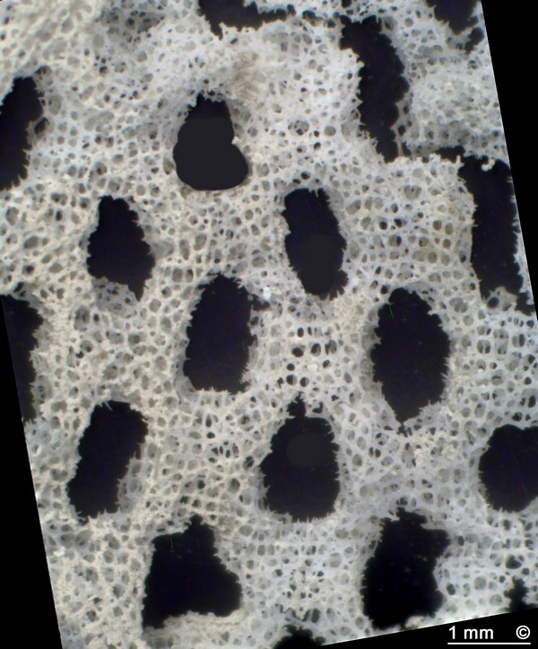
Figure 3 - Polyopesia angustata. Teutonia Nord, Misburg.
Stem section with roots.


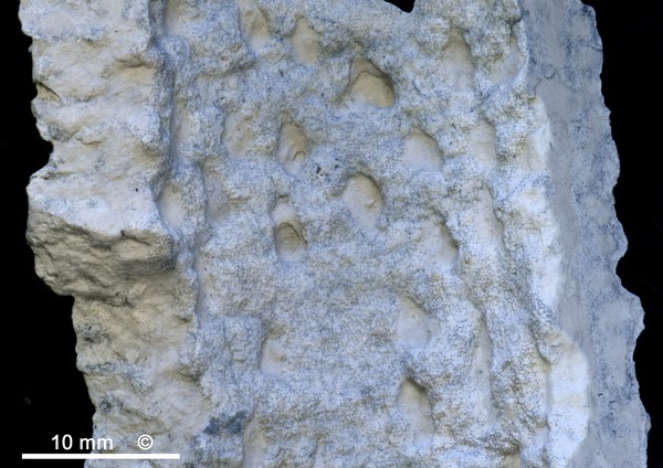
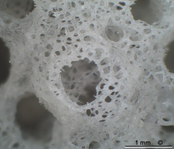
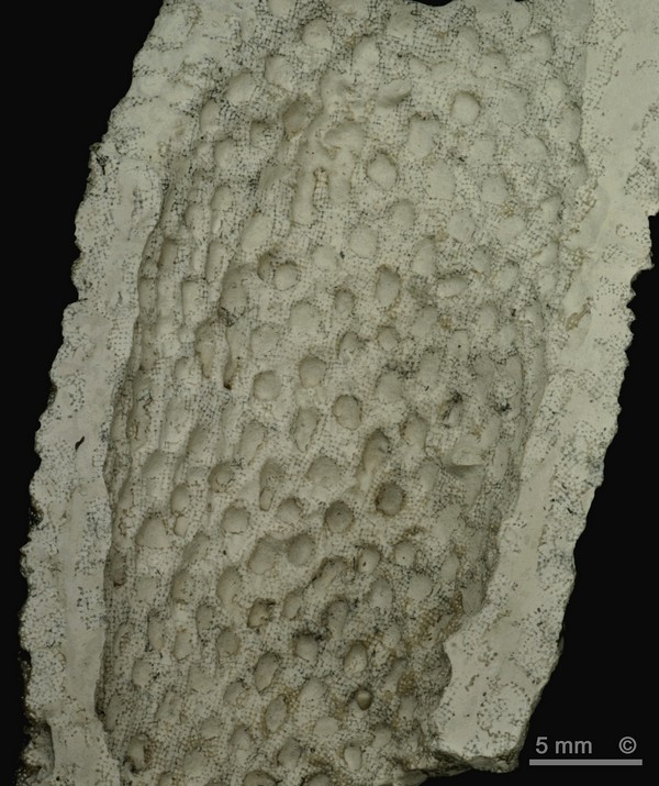
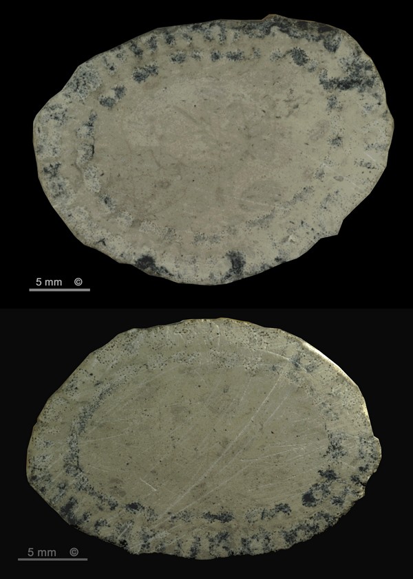
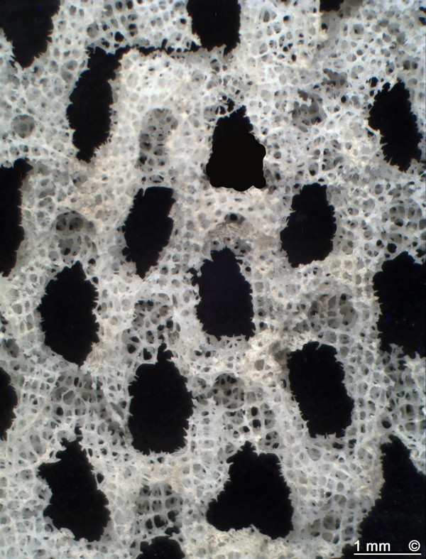
Figure 4 - Polyopesia angustata. Alemannia, Höver
Gastral side with postica.


Synonyms:

Hexactinella angustata Schrammen 1912
Hexactinella radiciformis Schrammen 1902

The nomenclature of the genus Polyopesia is confused and its taxonomic position is subject to dispute. (See below.)


Occurence:

Alemannia, Höver, Lower Campanian (senonensis zone). Rare.

Teutonia, Misburg, Upper Campanian (spiniger zone). Rare.


Figure 1 shows the most complete specimen of Polyopesia angustata found to date. Figure 2 shows a top-section with a growth margin, and Figure 3 shows a well preserved lower section with root stubs.

Considering all available material, typical individuals of Polyopesia angustata consist of a single tapering tube, some 200 to 300 mm tall and 20 to 40 mm wide, with a 6 to 10 mm thick wall. Senior individuals may develop a widened, trumpet-shaped top.

The roots are conspicuosly strong and are constructed from dictyonal hexactines. Near-end cross sections of the roots reveal several (5 to 10) longitudinal channels up to 3 mm wide.

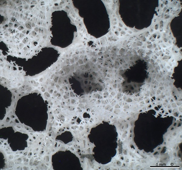
The dermal surface consists of many irregularly disposed protruding wart-like (external) postica, each 1.5 to 2 mm wide and separated from its neighbors by about the same distance (Figures 1 to 3). Ostia are arranged in depressed positions around the postica and are thus invisible in unetched specimens.

The gastral (atrial) side shows (internal) postica with quite large (4 to 5 mm) oval apertures, which are vaguely arranged in quincunx (Figure 4).

Figure 5 - Polyopesia angustata. Alemannia, Höver
(Etched specimen)


Figure 6 - Polyopesia angustata. Alemannia, Höver
Oblique view, emphasizing the protruding postica.
(Etched specimen)


Figure 7 - Polyopesia angustata. Alemannia, Höver
Two postica in the middle of seveal variably sized ostia.
(Etched specimen)


Figure 8 - Polyopesia angustata. Alemannia, Höver
Detailed view of lower right postica in previous figure.
(Etched specimen)


Figure 9 - Polyopesia angustata. Alemannia, Höver
Internal view, dermal side, showing euretoid dictyonal
framework (top left corner), free thorn-like hexactine arms
inside of aporhyses, and reinforced dermal hexactins
around ostia.
(Etched specimen)

Etched samples of Polyopesia angustata (Figures 5 and 6) reveal the ostia in the depressed positions between the wart-like postica.

Figure 7 is a photomicrograph of an etched sample, showing two wart-like raised postica with constricted apertures in the middle of several ostia with variablly sized apertures. Figure 8 is a detail of Figure 7. Note the silica mantled hexactins which constitute a dermal cortex.

Figure 9 (photomicrograph, etched sample) is an internal view of the dermal side, showing dyctional framework. Free thorn-like hexactine arms make up the inside of the aporhyses leading to the external postica. Note the reinforced dermal hexactins which also form the apertures if the ostia.


Polyopesia laevis

Schrammen 1912

Figure 1 - Hexactinella laevis, Teutonia Süd, Misburg
Large specimen with mushroom-shaped top.


Figure 2 - Polyopesia laevis, Allemania, Höver
Tubular specimen with growth margin.


Figure 3 - Polyopesia laevis, Teutonia Nord, Misburg
Senior individual with fully developed mushroom-shaped top.


Figure 4 - Polyopesia laevis, Alemannia, Höver
Funnel-shaped example.


Figure 5 - Polyopesia laevis, Alemannia, Höver
Trumpet-shaped top, dermal and gastral views.


Figure 6 - Polyopesia laevis, Teutonia Nord, Misburg
Senior individual with mushroom-shaped top and typical dermal surface structure.


Figure 7 - Polyopesia laevis, Teutonia Nord, Misburg
Longitudinal section, showing array of gastral postica and internal structure of walls.


Figure 8 - Polyopesia laevis, Teutonia Nord, Misburg
(Details of image above.)
Longitudinal sections through wall, showing sediment-filled longitudinal channels.


Figure 9 - Polyopesia laevis, Teutonia Nord, Misburg.
Cross sections, showing sediment-filled Channel system inside walls.


Synonyms:

Hexactinella laevis Schrammen 1912


Occurence:

Alemannia, Höver, Lower Campanian (senonensis zone). Rare.

Teutonia, Misburg, Upper Campanian (spiniger zone). Rare.


Polyopesia laevis forms straight to slightly widening tubes that may exceed 300 mm in height and 50 mm in diameter, as well as funnels. Senior individuals show a widened trumpet- to mushroom-shaped top. Supported by several strong divergent roots made up of euretoid dictyonal hexactins.

Compared to Polyopesia angustata, the dermal surface structure of un-etched Polyopesia laevis looks quite different. The surface of Polyopesia laevis has a more flat appearance since the postica are not as strongly protruding. Therefore, the large (2 to 3 mm) ostia of Polyopesia laevis become visible while the smaller dermal postica are less conspicuos.

On both surfaces, gastral and dermal, the hexactins are reinforced by a silica mantle, thus forming a rather strong cortex.

Figure 1 shows a tall and probably senior example of Polyopesia laevis, with a partially preserved mushroom-shaped top and strong roots.

The tubular sample shown in Figure 2 has not reached its full size and diaplays a growth margin at its top, whereas the senior specimen in Figure 3 shows a well developed mushroom-shaped top.

Figures 4 and 5 show more funnel-shaped individuals.

The typical surface structure of (un-etched) Polyopesia laevis can be seen in Figure 6.

Figure 7 reveals the gastral surface structure of Polyopesia laevis, with a more or less regular array of postica arranged in quincunx. The (gastral) postica are smaller (1 to 1.5 mm) and more numerous compared to those of Polyopesia angustata.

Figures 8 and 9 are longitudinal or cross sections through tubular sections of Polyopesia angustata. The sections reveal a complicated internal wall structure, with a system of essentially longitudinal channels. This channel system effectively separates the wall into an inner (gastral) and an outer (dermal) layer, with few points of contact. (This is the reason why coherent skeletal samples through the entire wall thickness cannot be obtained by acid leaching.) The chanellization system is considered equivalent to a system of schizorhyses, as described by Reiswig (2002, in Hooper and van Soest: Systema Porifera).

Figure 10 - Polyopesia laevis, Alemannia, Höver
(etched specimen). Note larger ostia in depressed cavities
between ridges, and smaller postica astride the ridges.


Figure 11 - Polyopesia laevis, (etched specimen).
Inside of dermal surface, showing dictyonal framework
between ostia and postica.


Figure 12 - Polyopesia laevis, (etched specimen).
Inside of dermal surface, with longitudinal (north-south)
ridges of dictyonine framework between ostia.
Note chambers, some with postica, between longitudinal ridges.


Figure 13 - Polyopesia laevis. Detail of previous figure.
Note spur-like free hexactin arms protruding into chambers
and channels.


Figure 14 - Polyopesia laevis, (etched specimen).
Gastral surface, with postica arranged in quincunx.


Figure 15 - Polyopesia laevis, (etched specimen).
Inside of gastral surface, with postica arranged in quincunx.


Figure 16 - Polyopesia laevis, (etched specimen).
Detail of euretoid dictyonine framework and free
hexactin arms.

Etched specimen of Polyopesia laevis (Figure 10), showing macroscopic structure of the dermal side, with larger ostia in depressed cavities between ridges, and smaller postica astride the ridges. The disposition of the dermal ostia appears more or less random. In other cases, a weak in quincunx arrangement of the dermal ostia may be recognizable.

Figures 11 through 13 give an impression of the internal wall structure (dermal layer) and the schizorhysal channelization. Figures 14 and 15 show aspects of the gastral (atrial) layer of the "double wall". In figure 15, note chambers adjacent to postica in longitudinal (north-south) direction, which are part of the schizorhysal channelsystem.

Figure 16 is a detailed view at the dictyonal framework. Note predominantly four-sided meshes. The nodes of the hexactins are not swollen in this case. However, in other samples more or less distinctly swollen nodes do occur. This feature therefore seems indistinctive for the species.


Note:

For reasons of consistency, the author adheres to the taxonomic classification by Reid (2004) for the moment and uses the designation "Polyopesia" rather than "Hexactinella".

Schrammen (1902) errected the new genus Polyopesia, with the two new species Polyopesia angustata (Plate II, Fig 1) and Polyopesia radiciformis (Plate III, Fig. 4). At that time, he tentatively placed Polyopesia into the family Craticularidae RAUFF 1893.

In his later work, Schrammen (1912) separated the tube- to funnel-shaped Polyopesia-species from the branched to anastomosed tubular Tretodyctium-species, moved the former into the family Tretocalycidae SCHULZE 1904 (syn. Tretodictyidae SCHULZE 1886) and the emended genus Hexactinella CARTER and consequently renamed his former Polyopesia into Hexactinella.

At the same time, Schrammen (1912) described "Hexactinella laevis" as a new species and synonymized Polyopesia radiciformis with Polyopesia angustata.

Reid (2004), without provision of arguments, considered Schrammen's (1912) identification of Polyopesia with Carter's Hexactinella to be wrong and transferred Polyopesia to the family Cribrospongiidae ROEMER 1864.


Author's comment:

The evidence provided here by the present author suggests that Schrammen's (1912) taxonomic classification was fully justified, and that the name Hexactinella should be used instead of Polyopesia. Polyopesia angustata and Polyopesia laevis should be regarded members of the family Tretodictyidae SCHULZE 1886.


BACK
HOME