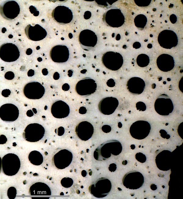
Figure 1 - Pleurope lacunosa. Alemannia, Höver.
Most complete specimen of its kind known to date.
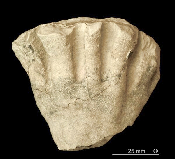
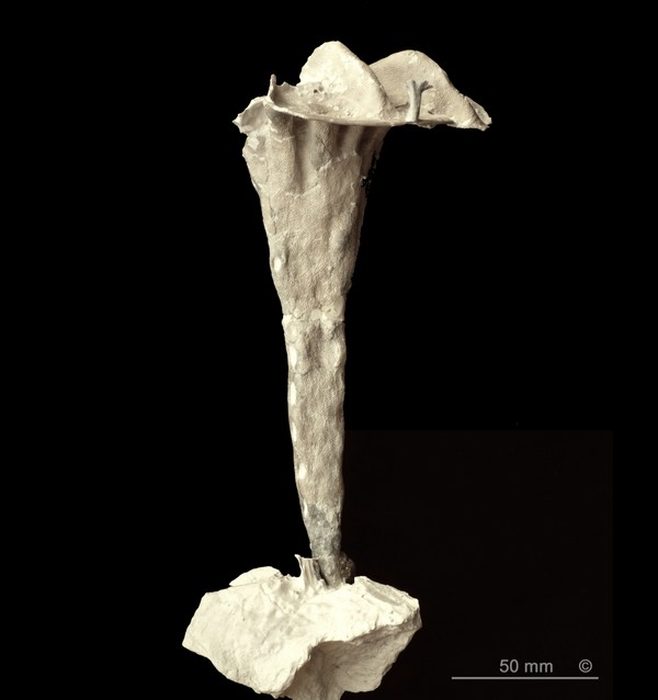
Figure 2 - Pleurope lacunosa. Alemannia, Höver.
Transition from stem to plicated funnel.
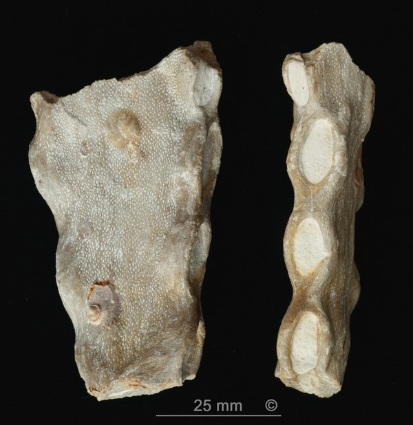
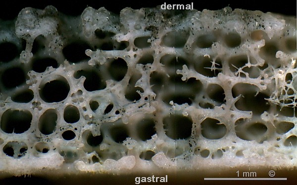
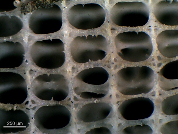
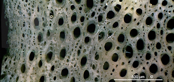
Figure 3 - Pleurope lacunosa. Oberg, near Peine.
Two views of a typical stem section.
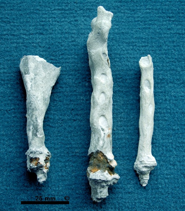
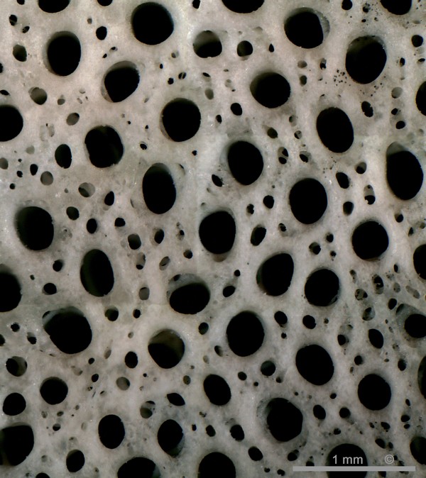
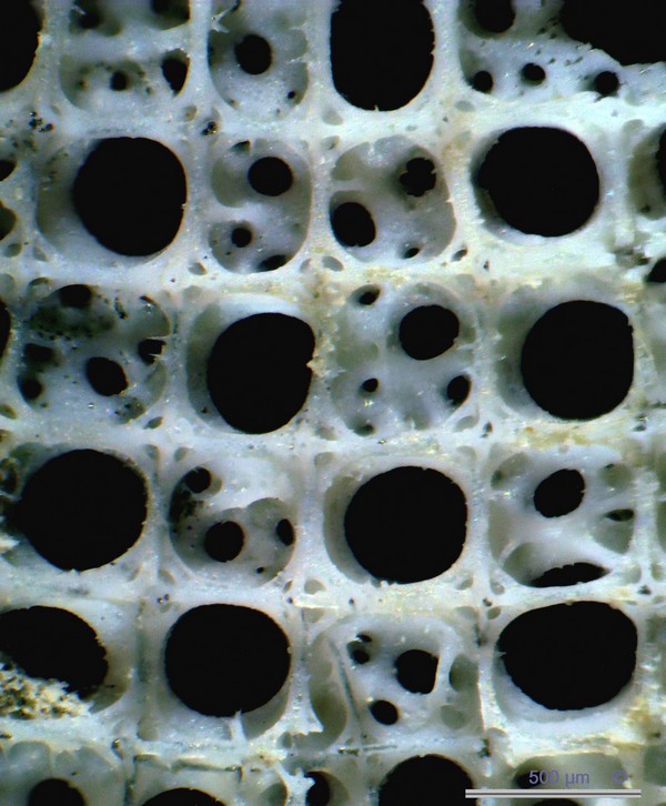
Figure 4 - Pleurope lacunosa. Alemannia, Höver.
Typical stem sections with holdfasts.
Synonyms:
Pleurostoma lacunosum Roemer 1841
Occurence:
Teutonia, Misburg, Upper Campanian (spiniger zone). Rare.
Alemannia, Höver, Lower Campanian (senonensis zone). Rare.
Previously, only the stems of Pleurope lacunosa were known and were actually mistaken for the sponge itself. The specimen shown here in Figure 1, although incomplete, reveals the true nature of Pleurope lacunosa.
The height of the specimen shown in Figure 1 is 210 mm, the original diameter of the funnel was approximately 120 mm; the ribs are approximately 6 to 7 mm thick; the wall thickness is 1 mm at the outer rim.
Pleurope lacunosa has a flat, scabbard-like stem with large oval openings (parietal oscula) along its narrow margins. However, approximately at half-height of the sponge, the stem widens to form a longitudinally folded funnel with a trumpet-shaped top (Figures 1 through 3). The inward-facing longitudinal folds of the lower funnel section turn into a set of pronounced radiating arched ribs. The outward-facing longitudinal folds of the lower funnel section grade into the flat sectors between the ribs.
Pleurope lacunosa is not known to form branching roots. The basal parts shown here apparently penetrated other objects to obtain some anchorage. However, the specimen shown in Figure 1 reveals tufts of siliceous fibres radiating laterally from the stem base and erecting at some distance (10 to 20 mm) from it.