cf. Phrissospongia glandiformis
Moret 1926
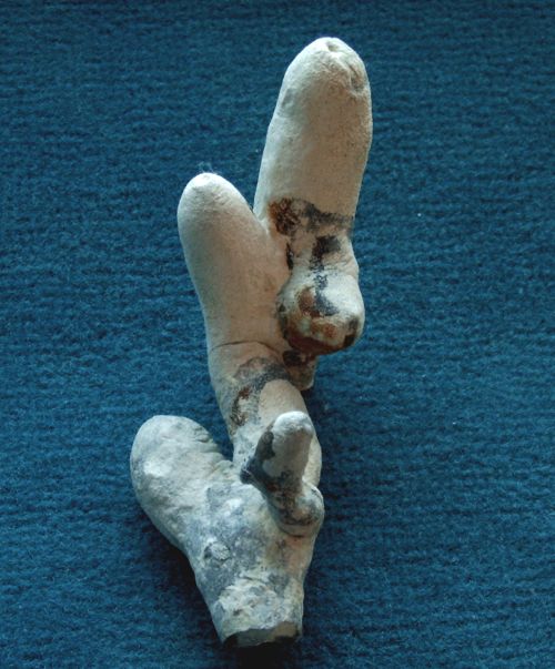
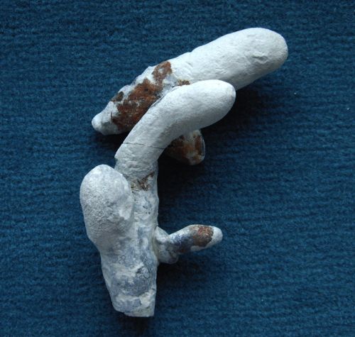
Phrissospongia glandiformis has been described by Moret (1926) from Santonian deposits near Saint Cyr, France. The specimen presented here in two different views is very closely related, but at the same time exhibits subtle differences in the sculpturing of its dermal scleres. The species has not been reported from Misburg or Höver previously, and to the author's knowledge neither from any other locality in Germany.
Moret's (1926) Phrissospongia glandiformis forms cylindrical individuals, which occur frequently as v-shaped "twins". The sponge has a deep but narrow, tube-shaped paragaster. Around its osculum, radial grooves are common. The sponge from Höver matches this macroscopic description completely.
Moret's Phrissospongia glandiformis has a very dense skeleton composed of predominantly tripodal dicranoclone desmas and subordinate megarhizoclonids. The dermal scleres are long-shafted dichotriaenes with characteristic short spines on the cladome. (See figure below, taken from Moret (1926).)
By comparison, the sculpturing of the dermal scleres of the specimen from Höver is slightly different (see below). In the unlikely case that Moret's (1926) description and his drawings of the dichotriaenes of Phrissospongia glandiformis are a bit schematic, then the specimens from Saint Cyr and Höver could belong to the same species. More likely however, we are dealing with a separate and new species closely related to Phrissospongia glandiformis.
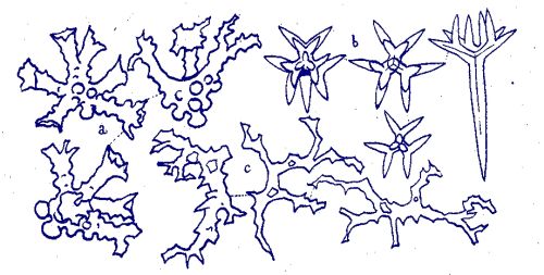
Spiculation of Phrissospongia glandiformis, as sketched by Moret (1926).
(a) dicranoclones
(b) sculptured dichotriaenes
(c) megarhizoclonids
_spic_001.jpg)
_spic_004.jpg)
_spic_006.jpg)
_spic_002.jpg)
_spic_003.jpg)
Spiculation of cf. Phrissospongia glandiformis from Höver. - Photomicrographs of an etched fragment of the above shown specimen
The first photomicrograph shows two megarhizoclonids in cavities of the dicranoclone skeleton. A dermal dichotriaene is visible in the top left corner.
The second picture shows the normal dicranoclone skeleton and the transition to the dermal layer (bottom of image) composed of sculptured dichotriaenes.
The third picture is a closer view of the dermal layer, composed of tiny, sculptured dichotriaenes.
The fourth picture is a close-up view of a megarhizolone surrounded by dicranoclones.
The fifth image shows a dermal dichotriaene in situ.
Isolation of individual scleres proved very difficult, due to the densely intergrown and stable nature of the skeleton. Even completely acid-leached samples do not disintegrate. The photographic equipment had to be pushed to its limits since the scleres of cf. Phrissospongia glandiformis are extremely small by comparison to almost all other sponges.
_Plate001.jpg)
Isolated dermal dichotriaenes of cf. Phrissospongia glandiformis from Höver.
Typically, the forked clads of the triaenes show short side branches near their tips and within the fork plane, making them look like snow crystals. Additionally, there are wart-like knobs on the outward facing sides of the clads.
The bottom right dichotriaene is shown from its underside, shaft (rhabdome) broken off. Notice black lines in rhabdome and clads which correspond to former axial filaments.
_Plate002.jpg)
Isolated dicranoclones and megarhizoclonids (bottom right) of cf. Phrissospongia glandiformis from Höver.
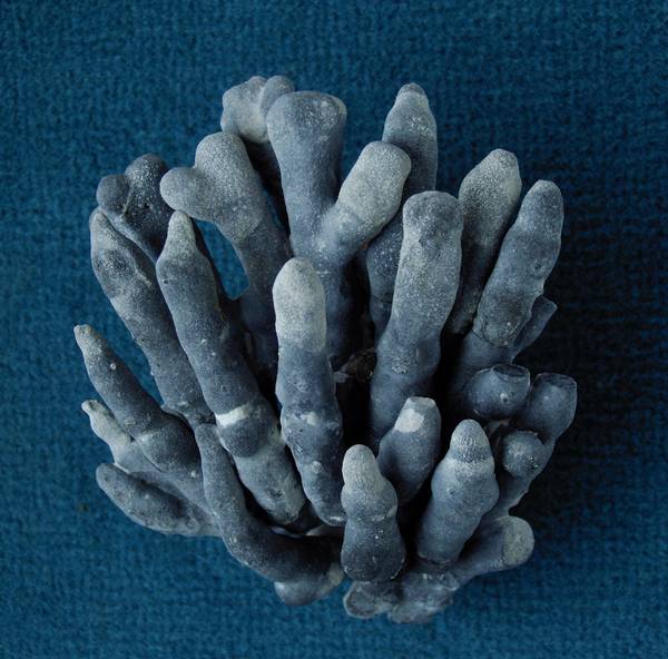
A larger specimen of cf. Phrissospongia glandiformis with numerous branches.
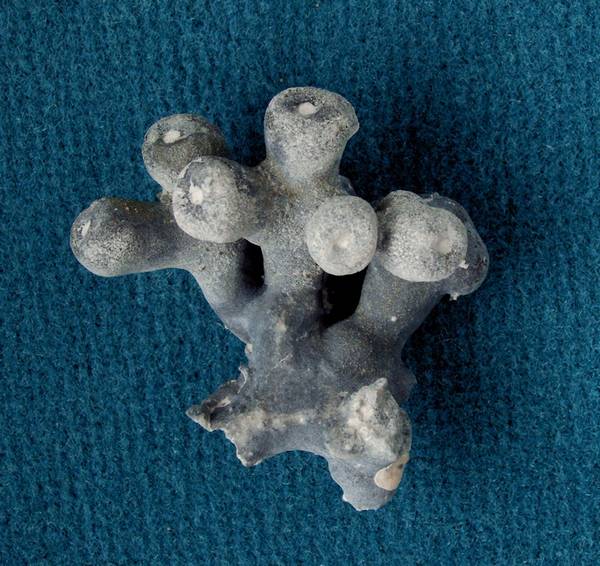
Isolated branch of cf. Phrissospongia glandiformis.
Notice symmetrical branching, swollen branch ends, and narrow oscula.