
Phalangium scytaliforme
Schrammen 1910
Phalangium scytaliforme was reported and described from Misburg by Schrammen (1910). He distinguished this species from Phalangium cylindratum (see below) on the basis of its habit: Phalangium scytaliforme is more stubby, with aspect ratios of approximately 1:3 whereas Phalangium cylindratum is more slender. However, the latter species was reported only from Groß Heere, not from Misburg.
As more specimens accumulated, it became obvious that most specimens from Misburg are also Phalangium cylindratum, and they were renamed accordingly. However, in the author's opinion there is little justification for the distinction of the two species.

Phalangium cylindratum
Schrammen 1910
The present example of Phalangium cylindratum is of cylindrical or club-shape, with a pointed top and a paragaster aperture (9 mm) on its apex. The lower end is broken off, but a number of roots emerging from (and below) a ring-like bulge are preserved.

Longitudinal section of Phalangium cylindratum. Notice the deep paragaster and straight, inclined epirhysal and aporhysal canals.

... Another example of Phalangium cylindratum. The specimen was originally mistaken for Pachinion cylindricum, due to the well developed cortex with abundant dermal dichotriaenes in the root region, which is more typical for Pachinion.
The unbranched roots emerging from the side rather than the bottom of the sponge appear to be characteristic for Phalangium cylindratum.

The last example shows a Phalangium cylindratum with a more truncated top. The specimen has toppled at some stage and subsequently developed small roots where lying on the ground. The subsequent growth direction has been diverted upward.
")
Skeletal organization of Phalangium, with dicranoclones, and with dermal dichotriaenes forming a cortex. After Moret (1926).
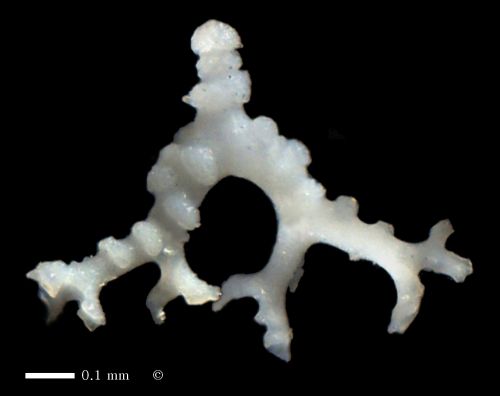
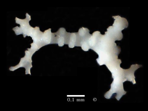
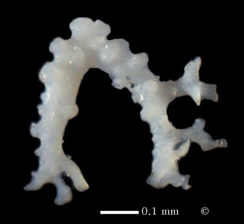
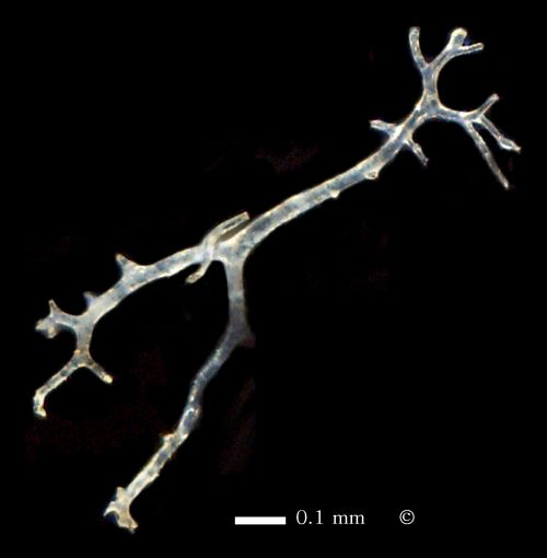
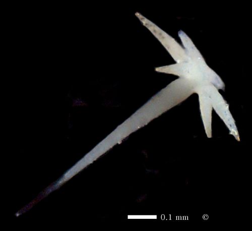
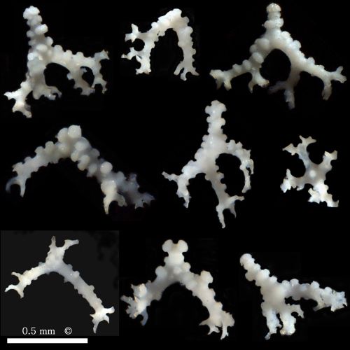
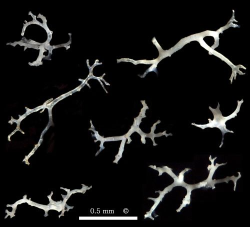
Scleres of Phalangium cylindratum.
The first three images are photomicrographs of dicranoclones. Notice the wart-like sculpturing.
The next image shows a rhizoclonide. Such rhizoclonides are quite abundant constituents of the skeleton and are larger, but much thinner than the dicranoclones.
The last photomicrograph shows a typical dermal dichotriaene. The dichotriaenes may also have cladomes distinctly shorter than in this example.
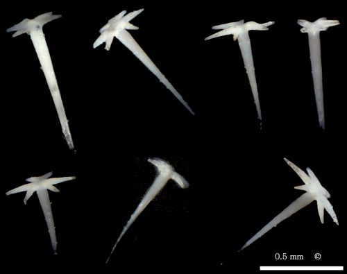
A collection of typical
- dicranoclones,
- pseudorhizoclones and
- dichotriaenes.