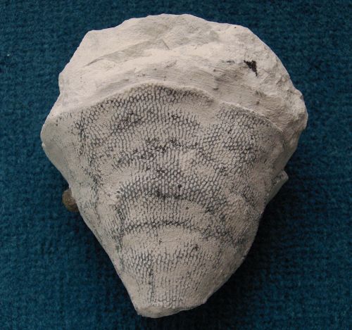
Napaea striata
Schrammen 1902
Napaea striata is probably absent from the Upper Campanian beds of Misburg. It is a moderately rare species in the Lower Campanian of Höver.
Napaea striata forms thin-walled (less than 2 mm), obconical funnels, rather similar to Sporadoscinia venosa, but its ostia and postica are aligned in longitudinal rows, giving it a striated appearance. The funnel is pendunculated, and its margin is usually straight, but may also be trumpet-shaped.
The ostia are small (less than 1 mm), slightly oval in longitudinal direction, and are generally arranged in quincunx.
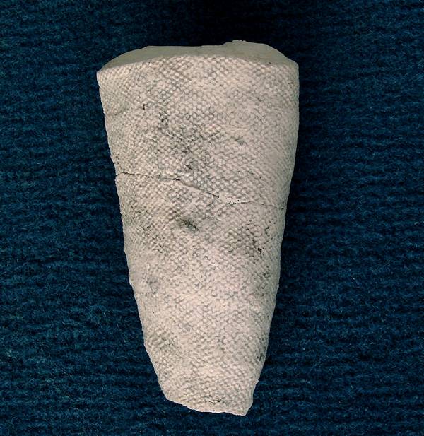
The basket of may also attain steep obconical shapes.
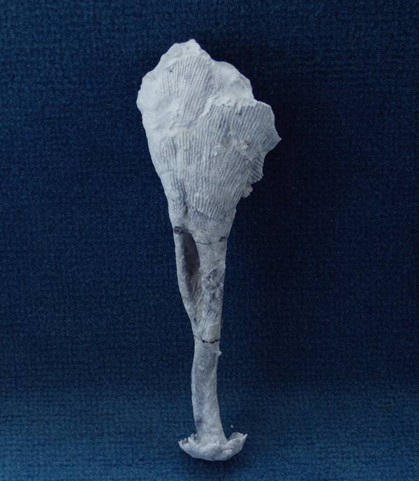
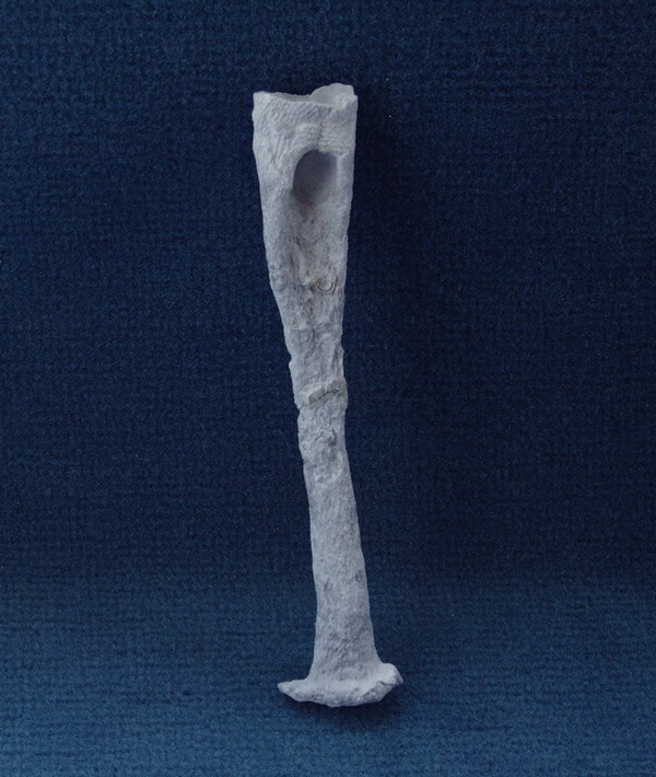
The next two images show stems and basal plates of Napaea striata.
Notice the large holes in the uppermost stem sections. These holes are connected to the bottom of the paragaster and probably served to prevent accumulation of debris inside the funnel.
Notice also the basal plates which are a special kind of anchorage typical for Napaea striata.
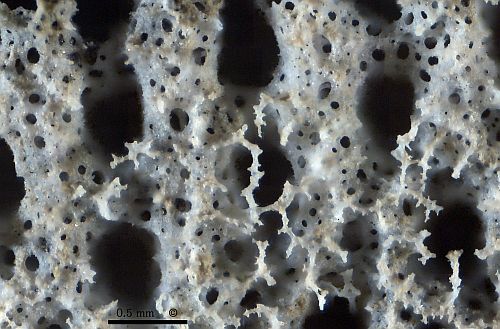
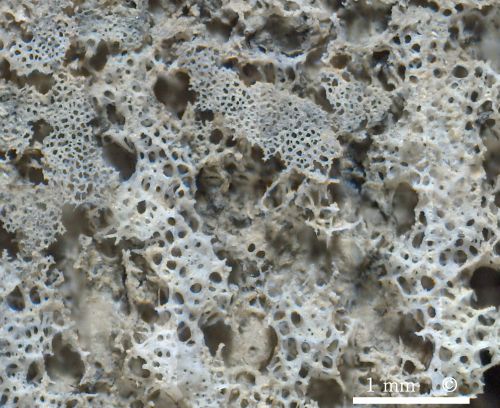
Photomicrographs of etched fragments of Napaea striata -- dermal and gastral surface.
The dermal surface shows of a cortex consisting of a porous, siliceous membrane which forms the bridges between the inhalent pores (ostia). The membrane extends from the tangential rays of modified surface lychnisks. Above this layer is a second surficial layer (partially preserved in the image) of irregularly branching appendices which develop from the radial rays of the surface lychnisks. (specimen upright)
The gastral surface is difficult to expose. However, the image shows four vertical skeletal bands between rows of (sediment-filled) exhalent pores. In the upper half of the image, some relics of a very delicate surface layer are preserved. (specimen upright)
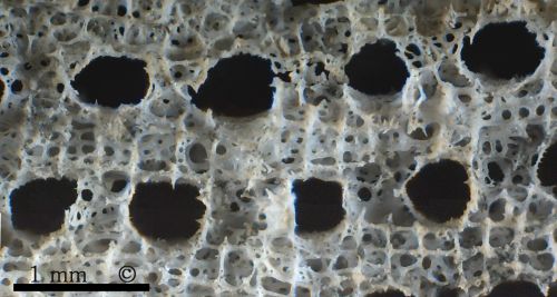
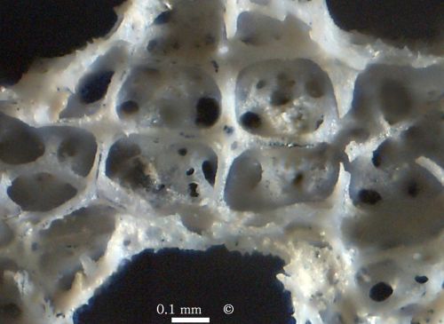
Photomicrographs of etched fragments of Napaea striata -- internal (parenchymal) skeleton.
The parenchymal skeleton consists of a fairly regular framework of lychnisks which surrounds the inhalent and exhalent channels. The image shows a view from inside the wall through the dermal pores. (specimen lying)
The last image shows a detailed view of the lychnisk-framework, with dermal membrane in the background. The lychnisk rays are generally smooth. Some axial canals in lychnisk-rays visible.